Gonioscopy
Through the use of a special lens, gonioscopy allows visualization and evaluation of the irido-corneal or drainage angle (where fluid produced inside the eye drains to the systemic circulation). Abnormalities of the drainage angle occur in some breeds and can be a predisposing factor for glaucoma.
Cytology
Corneal or conjunctival cytology can be helpful in the diagnosis of ocular disease. The ophthalmologist will collect cells from the surface of the eye and place them on a slide to be examined under a microscope.
Retinoscopy
Retinoscopy allows the refractive or dioptric state of the eye to be determined objectively. It can be used to define the normal, pathologic, and surgically induced refractive state of numerous domestic and exotic animal species.
ERG
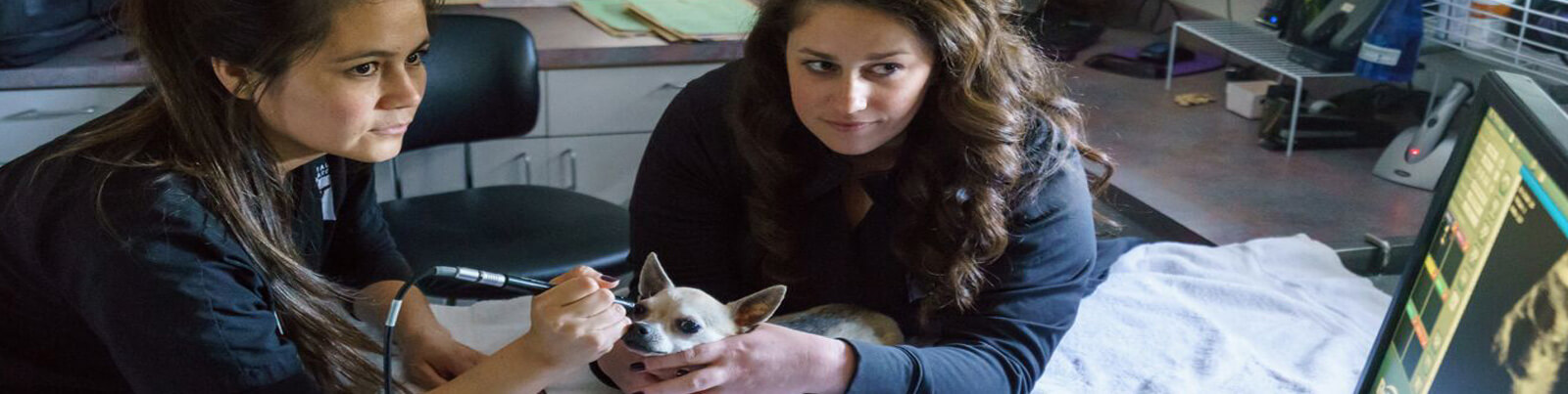
An ERG might be necessary to evaluate the function of the retina when the ophthalmologist suspects retinal disease or when the retina can not be visualized due to opacification of the eye (usually a cataract or corneal disease). An ERG records the total electrical response of the retina to a light stimulus by measuring the difference in potential between the retina and the cornea. An ERG is recorded by placing a special contact lens on the cornea and then flashing a series of bright lights into the eye. The patient will need mild sedation for this procedure. It can be performed at either of the Animal Eye Care clinics.
Samples
Diagnostic blood, urine or tissue samples can be collected and processed in-house or submitted to outside laboratories. These samples can be vital to reaching a rapid and accurate diagnosis of your pet’s condition. An accurate diagnosis assures proper treatment and gives your pet the best possible chance for a full recovery.
Ocular Ultrasound
Ocular ultrasound can be helpful in imaging ocular anatomy in cases where, due to opacities of the front part of the eye, it is not possible to visualize structures deeper inside the eye during the comprehensive exam. It helps to detect intraocular tumors, retinal detachments, lens luxation, or other abnormalities. Evaluating the orbit via ultrasound can help in the diagnosis of cysts masses or infections located behind the eye or “retrobulbar”. In most cases an ultrasound can be performed on short notice with the patient being awake. Both Animal Eye Care clinics are equipped with an ultrasound machine.
Radiograph
Skull radiographs can be helpful to detect fractures, tumors behind the eye (retrobulbar), or foreign bodies. Chest X-rays may be taken prior to general anesthesia in some animals, to rule out the presence of metastatic tumors, pneumonia or heart problems. Dacryocystorhinography is a technique in which a contrast medium is instilled into the nasolacrimal (tear) duct and radiographs are taken. This allows the localization of obstuctions of the nasolacrimal system. A board-certified radiologist may be consulted to assist in the interpretation of the findings.
Computerized Tomography
Computerized tomography is very helpful to detect lesions in areas that cannot be easily visualized. This technique is superior to MRI in regard to lesions of the bone, less sensitive for lesions of soft tissue or the nervous system. We cannot do this test at Animal Eye Care, but we can make recommendations about where to have it done. General anesthesia is necessary for CT scans.
MRI
The MRI provides images of structures deep inside the head that cannot be directly examined as the CT-scan. However, it is more sensitive for lesions within soft tissue or the nervous system. A CT-scan can be performed at our Fremont location. General anesthesia is necessary for the MRI.